|
Live-blood Analysis see
also: blood fluid flow
714-X
pleomorphism
ultramicroscopes
by Norman Allan
If you take a small drop of blood from a finger
tip and place it on a slide under a darkfield microscope you can watch patterns
in the live-blood. In standard microscopy light is shone directly through the
sample (specimen) and viewed against a bright field or background. However, we
cannot easily observe live material in this manner. Because a specimen must be
thin to be observed, it is also virtually transparent. Therefore we must stain
it to see it, and to do this we usually have to kill the specimen. This staining
process has become very sophisticated. There are hundreds of stains and hundreds
of questions you can ask about your specimen with these stains. And that, as far
as most microscopists are concerned, is that.
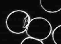
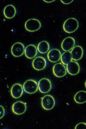
Red Blood Cells as seen in Darkfield
With darkfield microscopy, however, the procedure is crucially different.
The light does not travel directly through the specimen, but comes in from the
sides and only light which is reflected by the specimen is viewed and viewed against
a dark background, hence “darkfield microscopy�. In this way a highly contrasted
image is obtained and there is no need to use a stain. We can therefore watch
living material.
 Practitioners of live-blood analysis claim they can spot cancer and other
"degenerative immune system diseases" up to two years before they would otherwise
be detectable. Some practitioners say that they can see parasites. Almost everyone
who studies live-blood in detail comes up with their own story. Let me tell you
about a few of these, and then I'll tell you what I think I could and what I could
not see.
Practitioners of live-blood analysis claim they can spot cancer and other
"degenerative immune system diseases" up to two years before they would otherwise
be detectable. Some practitioners say that they can see parasites. Almost everyone
who studies live-blood in detail comes up with their own story. Let me tell you
about a few of these, and then I'll tell you what I think I could and what I could
not see.
Quebec scientist Gaston Naessens is a controversial figure
who invented a remarkable microscope using a complex UV light source. He called
it a "somatoscope" and with it he was able to examine living material at 30 times
the resolution available with conventional microscopes. This led him to the discovery
of what he calls "somatids". Somatid means "tiny bodies". According to Naessens
somatids are found in all living creatures. In a healthy organism somatids have
a simple three stage life cycle (somatid, spore, and double spore). In this "microcycle"
somatids are symbiotic: they perform essential functions in the regulation of
cell division, Naessens claims.
However, when we are ill the somatids
elaborate into a complex sixteen stage "macrocycle". The somatids of the macrocycle
are bacteria-like and fungus-like. This change of form is an example of what is
called "pleomorphism". Gaston Naessens' theory says that we see the
macrocycle in ill health or with impending illness. It is also claimed that with
the darkield microscope we may see the disease pattern in the blood up to two
years before a disease (for example cancer) manifests. The somatid cycle (discovered
by Naessens with the somatoscope and analyzed in culture) can be observed and
monitored with darkfield microscopy. We can use this to alert us to incipient
problems, or to monitor a patients response to therapies, both orthodox or alternative
therapies.
A second approach to live-blood analysis has been assembled
from many sources by Prof. Lida Mattman of the Wayne State University and her
colleague Dr. Phil Hockstra. Micro-organisms, when challenged, shed their cell
walls. While this leaves them less virulent, it also makes them less vulnerable.
Shedding their skins they lose most of the markers that identify them as foreign
bodies to our immune systems. They can also now change their shape - this simple
change of shape is also called pleomorphism - and all this means they can easily
invade and hide in the body's own cells.
In live-blood analysis as taught
by Dr. Hockstra attention is paid to the microbes, to the shape and activity of
the white blood cells (WBC) and red blood cells (RBC). By observing the red cells
we can tell a lot about the state of metabolism in general and of the liver particular.
Meanwhile, observing the white cells gives us a reading on the state of the immune
system, and the pattern of microbes tell us if disease is overwhelming the body's
defences. Note that while we call this "live-blood" analysis it is really dying
blood that we are observing. In a sense we are watching how quickly rot sets in
after we take the drop of blood out of the body and this tells us how much resilience
and vitality there is in the body - it is a measure of the body's health.
Toxoplasma gondii: an intracellular protazoan parasite
In both Naessens' and
Hockstra's systems the blood is observed for 30 minutes. There are however a large
number of practitioners who observe the live-blood for about five minutes with
a "phase-contrast" microscope and then make a "nutritional assesment". While one
can observe the red cells as well with a phase contrast condenser as with darkfield,
white cells, microbes, platelets and other blood elements are not as well visualized.
I also fear, personally, that much of this "nutritional analysis" is spurious.
But then I also disagree with Naessens and Hockstra on a number of points as well.
For instance, almost everyone puts undue emphasis on the fact that red
cells clump together in coin-like stacks (or rouleaux formation) and treat this
as pathological. In fact RBCs clump together in rouleaux
stacks wherever space allows them to. They do this, actually, to reduce the
viscosity of the blood and for the protection that the red cells receive by traveling
in these train-like processions.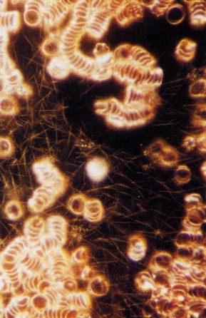 This
is a normal function of blood and, furthermore, its extent cannot be easily quantified
with live-blood work. (This is because we can not calculate exactly how thick
the sample is at any point.) The extent of this clumping, this rouleaux formation
can however be easily measured in standard blood work as sedimentation rate, or
SED rate. (If rouleaux formation is excessive the blood sediments settle more
quickly.) SED rate is a general indicator of inflammation. We generally expect
the SED rate to be elevated in inflammatory conditions such as arthritis. But
SED rate is a very general and imprecise measure, and medicine pays little attention
to it nowdays. This
is a normal function of blood and, furthermore, its extent cannot be easily quantified
with live-blood work. (This is because we can not calculate exactly how thick
the sample is at any point.) The extent of this clumping, this rouleaux formation
can however be easily measured in standard blood work as sedimentation rate, or
SED rate. (If rouleaux formation is excessive the blood sediments settle more
quickly.) SED rate is a general indicator of inflammation. We generally expect
the SED rate to be elevated in inflammatory conditions such as arthritis. But
SED rate is a very general and imprecise measure, and medicine pays little attention
to it nowdays.
Some microscopists claim that live-blood can be used
to diagnose for parasites. It is important to note that "parasites" are large
and generally live in the gut. In live-blood analysis we take a small drop of
blood, perhaps a billionth part. Your blood would have to be overrun with these
"giants" before we would see them. I've looked at hundreds of bloods and perhaps
I've seen a parasite once. We do, however, see a lot of break down products from
the blood cells. In particular the cell walls of the red cells will often unravel
just like a knitted jersey. Wiggly moving threads unravel from the red cell walls.
Their scientific name is myelin spindles, but some practitioners are calling
them "parasites". Just because it is moving does not mean that it is alive! At
microscopic dimensions it is normal for inanimate objects to move. This is caused
by thermal and by Brownian motion.
What do I see in live-blood analysis?
In my practice (1994 to 1999*) I have integrated several schools of darkfield
microscopy. I have been trained by Naessens step-sons in the somatidian analysis
and by Dr. Hockstra in his more conservative, but still controversial, approach.
It is my opinion that using these systems judiciously we can see, first, the health
of the "ground" or "terrain" - how healthy is the organism? are the red cells
well formed? We can see the pressure on the immune system - how stressed are the
white blood cells? Note, while we see the pressure in the system, we don't see
exactly when or how it is going to break down. We can see how close to being overwhelmed
the system may be - is the rot, the bacteria and fungus, taking over? Finally,
and most importantly, we can watch over time how a patient is responding to their
treatment.
There are also, of course, more specific things we can tell
at times. There are signs of free radical damage and oxidation http://www.kamagraoraljelly247.com/. And with yeasts,
candida, we can see some indication of how prevalent these are in the whole body.
Further, we always get a lot of information about the degree and the types of
anemia that may be present. But primarily I find darkfield live-blood analysis
a way of monitoring general health. It is particularly useful, as mentioned above,
in tracking patient's response over time to their treatments indicating at the
earliest moment a need to change a medication or therapy. In certain conditions,
especially cancer or other life threatening disease, where time is a crucial factor,
darkfield microscopy can possibly be of help in choosing and monitoring the effect
of therapy.
see also:
blood fluid flow
pleomorphism
714-X
note:
I did live-blood analysis from 1994 to 1999. However, I am no longer directly
involved in the field.
*I did "live-blood
analysis" from 1994 to 1999.
I have not done it since. It is a very interesting
phenomenon,
but at present nobody understands it well, and many of the people
claiming to
do live-blood analysis have a completely erroneous take on the
matter!
|